Diabetic retinopathy care in Suwanee, GA — early detection, expert monitoring

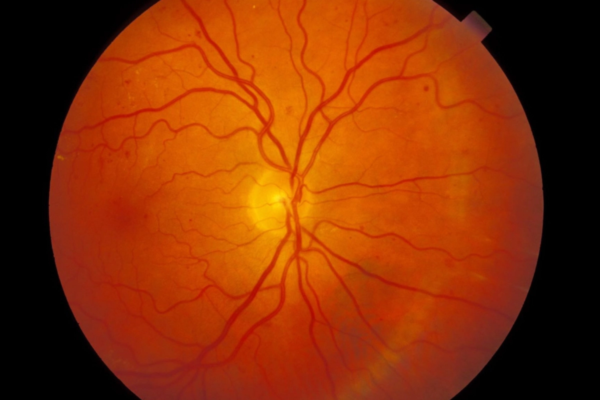
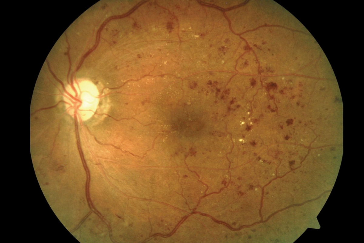
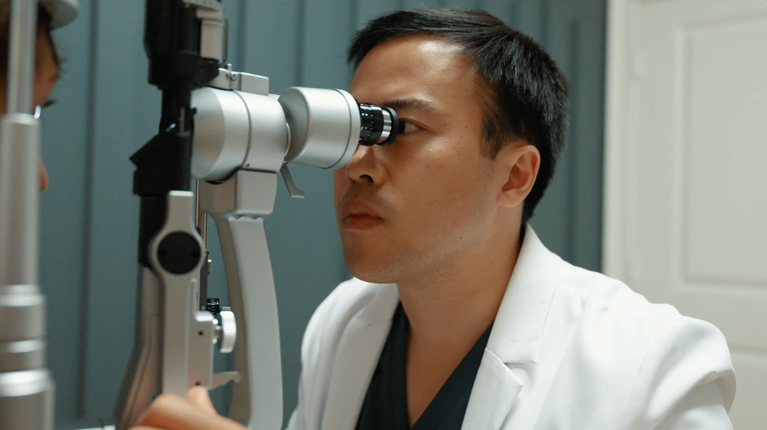
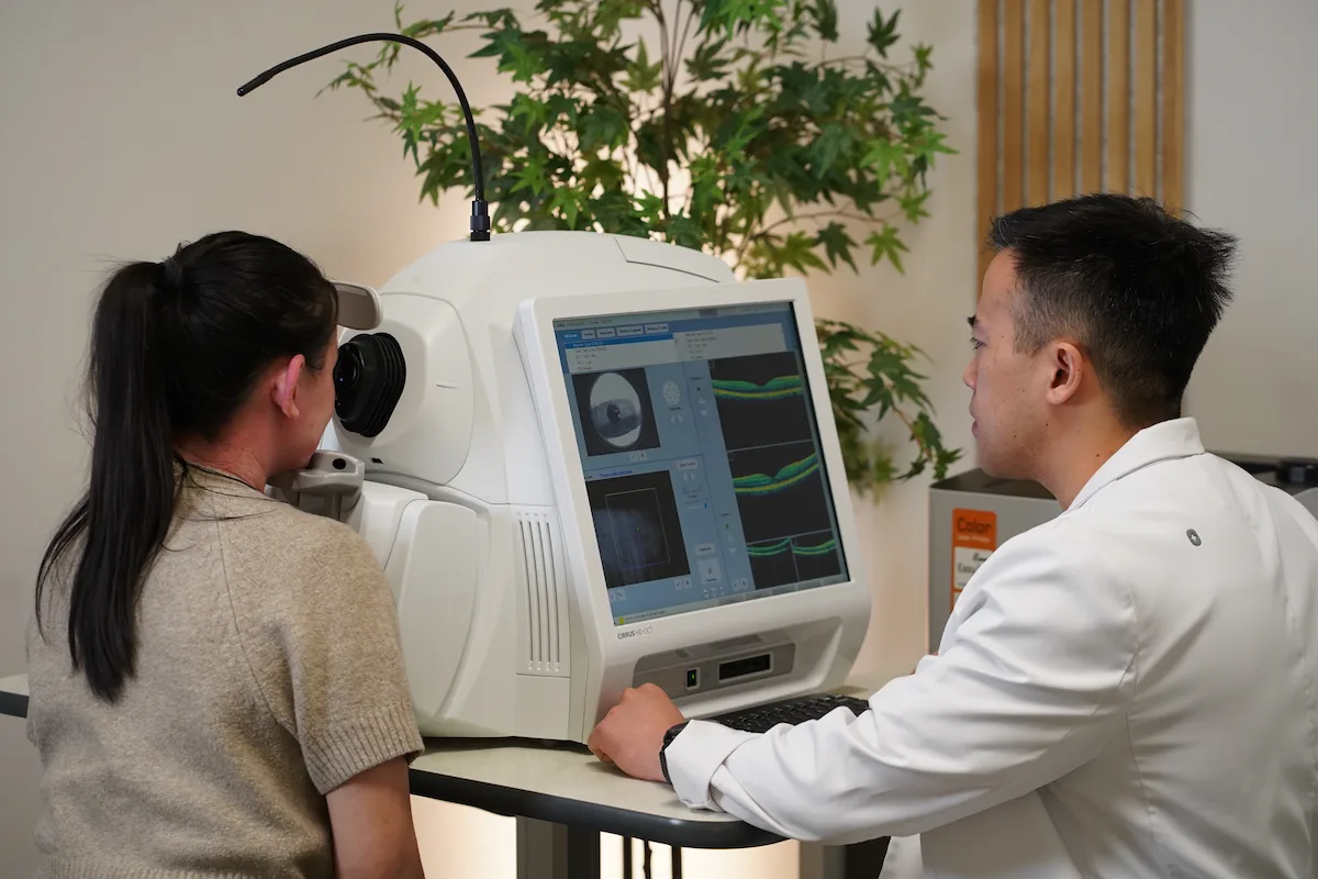
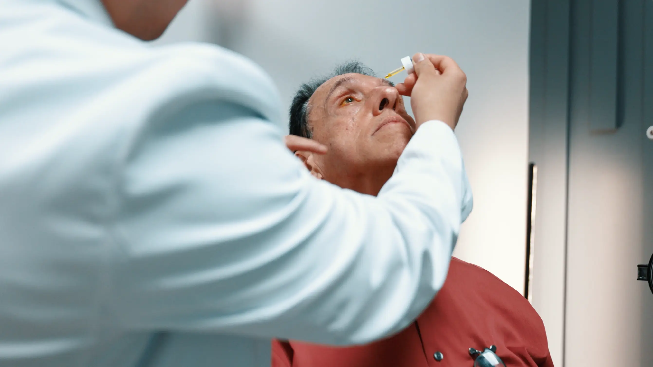
Diabetic retinopathy is the leading cause of preventable blindness in working-age adults in the United States. At Stratus Eye, Dr. Jeffrey Tran brings experience managing some of the most advanced cases in the country — and the same standard of care to every diabetic patient who walks through our door.
Dr. Jeffrey Tran performing macular degeneration evaluation at Stratus Eye in Suwanee, GA

What is diabetic retinopathy?

Diabetic retinopathy is damage to the blood vessels of the retina caused by chronically elevated blood sugar. Over time, high glucose levels weaken the walls of the tiny vessels that supply the retina — causing them to leak fluid, bleed, or grow abnormally. It is the leading cause of preventable blindness in working-age adults in the United States, and it affects roughly one in three people with diabetes to some degree.

What makes diabetic retinopathy particularly dangerous is its silence. In the early and intermediate stages, there are no symptoms. Vision can appear completely normal while significant damage is actively occurring behind the eye. By the time a patient notices something is wrong, the disease has often already reached an advanced stage — one that is far harder to treat and far more likely to result in permanent vision loss.

Every diabetic patient is at risk. Duration of diabetes and blood sugar control are the strongest predictors of progression — but retinopathy can develop even in patients who manage their diabetes carefully. Annual dilated eye exams are the only reliable way to detect it before it steals your vision.

Understanding the stages of diabetic retinopathy

Mild nonproliferative diabetic retinopathy — retinal photograph showing early stage changes at Stratus Eye in Suwanee, GA

Mild nonproliferative diabetic retinopathy (NPDR)

The earliest stage. Small areas of balloon-like swelling in the retinal blood vessels — called microaneurysms — begin to appear. There are no symptoms and vision remains normal. Most patients have no idea this is happening. A dilated exam is the only way to detect it.
Moderate to severe nonproliferative diabetic retinopathy — retinal photograph showing disease progression at Stratus Eye in Suwanee, GA

Moderate to severe NPDR

As the disease progresses, more blood vessels become blocked, depriving the retina of its blood supply. The retina responds by signaling for new blood vessel growth — a dangerous turning point. Vision may still appear normal, but the window for the most effective intervention is actively narrowing.
roliferative diabetic retinopathy — advanced stage retinal photograph at Stratus Eye in Suwanee, GA

Proliferative diabetic retinopathy (PDR)

The most advanced stage. Fragile, abnormal new blood vessels grow across the retina and into the vitreous. These vessels bleed easily, causing floaters, sudden vision loss, and retinal detachment. PDR can also trigger neovascular glaucoma, which is one of the most aggressive and difficult-to-treat forms of glaucoma. This occurs when abnormal vessels invade the eye's drainage angle.

While there is no cure, progression can be slowed with monitoring, lifestyle modifications, and in some cases nutritional supplementation based on the AREDS2 formula.

Diabetic macular edema (DME)

DME can occur at any stage of diabetic retinopathy. Fluid leaks from damaged blood vessels into the macula — the center of the retina responsible for sharp, detailed vision — causing it to swell.

It is the most common cause of vision loss in diabetic patients and requires prompt evaluation and coordinated care with a retina specialist.
OCT scan showing diabetic macular edema — fluid accumulation in the macula at Stratus Eye in Suwanee, GA

How do you know if you have diabetic retinopathy?

01
No symptoms at all
This is the most dangerous presentation. Mild, moderate, and even severe NPDR frequently cause zero noticeable vision changes. The damage is progressing — you just can't feel it. A dilated exam is the only way to know where you stand.
02
Floaters or dark spots
As diabetic retinopathy advances, fragile new blood vessels can bleed into the vitreous — the gel-like fluid inside the eye. This causes sudden floaters, dark streaks, or shadowy spots in your vision. This is a sign of advanced disease and requires immediate evaluation.
03
Blurry or fluctuating vision
Fluid leaking into the macula from damaged blood vessels causes swelling that blurs central vision. This may fluctuate throughout the day depending on blood sugar levels — often dismissed as a glasses prescription issue when diabetic macular edema is the actual cause.
04
Sudden vision loss
Bleeding from abnormal new blood vessels, retinal detachment, or neovascular glaucoma can each cause sudden, severe vision loss. These are ophthalmic emergencies. If you are diabetic and experience sudden vision changes, call immediately — do not wait for a scheduled appointment.
If you have diabetes and haven't had a dilated eye exam in the past year, that's the right next step — regardless of whether your vision feels normal.
Arrow
Schedule an Appointment

What causes diabetic retinopathy?

The root cause is chronically elevated blood sugar. Over time, high glucose levels damage the walls of the small blood vessels that supply the retina — causing them to weaken, leak, and eventually close off entirely. The retina, deprived of oxygen and nutrients, responds by triggering the growth of new abnormal blood vessels. Those vessels are fragile, prone to bleeding, and the primary driver of vision-threatening complications.

Several factors accelerate this process: poorly controlled blood sugar, high blood pressure, high cholesterol, pregnancy, and longer duration of diabetes all increase both the risk of developing retinopathy and the speed at which it progresses. Type 1 and Type 2 diabetics are both at risk — though the timeline and presentation can differ.

Dr. Tran trained at Grady Memorial Hospital in Atlanta and Ben Taub General Hospital in Houston — two of the busiest safety-net hospitals in the United States, serving patient populations with some of the highest rates of advanced, undertreated diabetic retinopathy in the country. That clinical experience means he has evaluated and managed the full spectrum of this disease — from the earliest microaneurysms to sight-threatening PDR — and brings that standard of care to every diabetic patient at Stratus Eye.
Dr. Jeffrey Tran performing a diabetic retinopathy evaluation at Stratus Eye in Suwanee, GA

How we evaluate and monitor diabetic retinopathy

A thorough exam today is the only reliable protection against vision loss tomorrow.
Step 1

Your comprehensive diabetic eye exam

Your first visit includes a dilated fundus exam and OCT imaging — giving Dr. Tran a direct, detailed view of your retina, macula, and blood vessels. Every finding is reviewed with you personally. You'll leave knowing exactly what stage of retinopathy you have, whether DME is present, and what needs to happen next.
Step 2

Your monitoring plan

Based on your exam findings, Dr. Tran establishes a monitoring schedule matched to your stage of disease. Mild NPDR typically warrants annual exams. Moderate to severe NPDR requires closer follow-up every 3–6 months. Any new symptoms between visits — floaters, sudden blurring, vision changes — are evaluated immediately, not at your next scheduled appointment.
Step 3

Coordinated care when you need it

When retinal intervention is needed — laser photocoagulation, anti-VEGF injections, or vitreoretinal surgery — Dr. Tran coordinates your referral directly to a trusted retina specialist. You are not handed a name and sent on your way. Stratus Eye remains your primary eye care home base, managing your annual exams, monitoring disease stability, and staying connected to your broader diabetes care team.
ABOVE THE STANDARD
How is diabetic retinopathy care different at Stratus Eye?

Diagnosis is only as good as the technology and the hands behind it

Dilated fundus exam

Dilation allows Dr. Tran to examine the full retina directly — evaluating blood vessel integrity, identifying microaneurysms, hemorrhages, and neovascularization, and assessing the optic nerve in the same visit. No referral needed, no separate appointment. Every diabetic patient receives a dilated exam at every comprehensive visit.

Dr. Jeffrey Tran performing a diabetic retinopathy evaluation at Stratus Eye in Suwanee, GA

Optical Coherence Tomography (OCT)

The gold standard for detecting diabetic macular edema. OCT produces a precise, cross-sectional map of your macula — measuring retinal thickness, identifying fluid accumulation, and detecting structural changes that are completely invisible during a standard exam. It's how we catch DME early, before it causes permanent central vision loss.

OCT machine used for diabetic macular edema detection at Stratus Eye in Suwanee, GA

Systemic risk factor review

Diabetic retinopathy doesn't exist in isolation. Blood sugar control, blood pressure, cholesterol, and medication adherence all directly influence how quickly the disease progresses. At every visit, Dr. Tran reviews your systemic risk factors alongside your retinal findings — and communicates with your primary care physician or endocrinologist when the clinical picture warrants it.

Diabetic retinopathy systemic risk factor review at Stratus Eye in Suwanee, GA

Intraocular pressure measurement

Advanced diabetic retinopathy carries a risk of neovascular glaucoma — one of the most aggressive forms of glaucoma, caused by abnormal blood vessels invading the eye's drainage angle. Intraocular pressure is measured at every visit for diabetic patients. Catching elevated pressure early is the difference between a manageable finding and a vision-threatening emergency.

Intraocular pressure measurement for diabetic retinopathy monitoring at Stratus Eye in Suwanee, GA

Frequently asked questions about diabetic retinopathy

What is diabetic retinopathy?

Arrow

Diabetic retinopathy is damage to the blood vessels of the retina caused by chronically elevated blood sugar. Over time, those vessels weaken, leak fluid, and can close off entirely — depriving the retina of oxygen and triggering the growth of abnormal new blood vessels. It is the leading cause of preventable blindness in working-age adults in the United States.

What are the stages of diabetic retinopathy?

Arrow

There are four main stages. Mild NPDR is the earliest, characterized by small microaneurysms in the retinal vessels. Moderate to severe NPDR involves more widespread vessel damage and blockage. Proliferative diabetic retinopathy (PDR) is the most advanced stage, marked by the growth of fragile new blood vessels that bleed easily and can cause retinal detachment. Diabetic macular edema can occur at any stage and is the most common cause of vision loss in diabetic patients.

What is diabetic macular edema?

Arrow

Diabetic macular edema occurs when fluid leaks from damaged retinal blood vessels into the macula — the central part of the retina responsible for sharp, detailed vision. The macula swells, causing blurry or distorted central vision. DME can develop at any stage of diabetic retinopathy and requires prompt evaluation and coordinated care with a retina specialist.

Can diabetic retinopathy cause blindness?

Arrow

Yes — it is the leading cause of preventable blindness in working-age adults in the United States. The critical word is preventable. When caught early and monitored appropriately, the risk of severe vision loss is dramatically reduced. The danger is that most patients have no symptoms until the disease is already advanced.

What is neovascular glaucoma?

Arrow

Neovascular glaucoma is a severe complication of advanced diabetic retinopathy. When abnormal blood vessels grow into the eye's drainage angle, they block fluid outflow and cause a rapid, dangerous rise in intraocular pressure. It is one of the most aggressive and difficult-to-treat forms of glaucoma. This is why intraocular pressure is measured at every diabetic eye exam at Stratus Eye — not just when symptoms appear.

How often should diabetic patients have eye exams?

Arrow

The American Academy of Ophthalmology recommends that all Type 1 diabetics have a comprehensive dilated eye exam within 5 years of diagnosis, and all Type 2 diabetics at the time of diagnosis. After that, the frequency depends on your stage of disease. Mild NPDR typically warrants annual exams. Moderate to severe NPDR requires follow-up every 3–6 months. Any new visual symptoms should be evaluated immediately.

Can diabetic retinopathy be reversed?

Arrow

Early diabetic retinopathy can partially stabilize or improve with better blood sugar control, blood pressure management, and lifestyle changes. However, structural damage that has already occurred cannot be fully reversed. This is why early detection matters so much — the earlier the disease is identified, the more of your vision there is to protect.

Does diabetic retinopathy always cause vision loss?

Arrow

Not always — and that's the point of monitoring. Many patients with early and intermediate NPDR never progress to vision-threatening disease, particularly with good systemic control and regular eye exams. The patients who lose vision are most often those who had no symptoms, assumed everything was fine, and skipped their annual exam.

What is the treatment for diabetic retinopathy?

Arrow

Treatment depends on the stage. Early NPDR is managed with monitoring and systemic risk factor control. Diabetic macular edema and PDR are treated by retina specialists with anti-VEGF injections, laser photocoagulation, or vitreoretinal surgery when needed. At Stratus Eye, Dr. Tran evaluates and monitors your disease and coordinates your referral directly when intervention is required.

I have diabetes but my vision seems fine. Do I still need an eye exam?

Arrow

Yes — this is exactly the patient who most needs a dilated exam. Normal vision does not mean healthy retinas. Significant diabetic retinopathy can be present and actively progressing with no symptoms whatsoever. The exam is not to check your glasses prescription — it is to evaluate your retinal blood vessels directly, which cannot be done any other way.

Can high blood pressure make diabetic retinopathy worse?

Arrow

Yes. High blood pressure accelerates retinal blood vessel damage independently of blood sugar levels. Patients with both poorly controlled diabetes and hypertension are at significantly higher risk of rapid progression to vision-threatening disease. Managing both conditions together is essential — and Dr. Tran reviews your systemic risk factors at every visit.

Does Stratus Eye treat diabetic retinopathy with injections or laser?

Arrow

Intravitreal injections and laser photocoagulation for advanced diabetic retinopathy are performed by retina specialists. At Stratus Eye, Dr. Tran provides comprehensive evaluation, monitoring, and risk factor management — and coordinates your care directly with a trusted retina specialist when intervention is needed. You remain our patient throughout.

SERVING NORTH ATLANTA

Diabetic retinopathy care for patients across North Atlanta

Stratus Eye is proud to serve diabetic patients from across the North Atlanta region. Whether you're coming from Suwanee, Johns Creek, Duluth, Alpharetta, Cumming, Buford, Sugar Hill, Gainesville, or Lawrenceville, Dr. Tran provides comprehensive diabetic retinopathy evaluations and ongoing monitoring — with the clinical experience and diagnostic technology to detect changes before they affect your vision. If you have diabetes and haven't had a dilated eye exam in the past year, this is the visit that matters.

TAKE THE NEXT STEP
Schedule a diabetic eye exam at Stratus Eye in Suwanee, GA — Dr. Jeffrey Tran

Ready to protect your vision before diabetes takes it?

A diabetic eye exam with Dr. Tran is the first step. We'll dilate your eyes, image your retina with OCT, measure your eye pressure, and give you a clear picture of where you stand — with no pressure and no obligation. Most patients leave with answers their primary care doctor has never given them. Diabetic retinopathy is preventable blindness. The exam is how you prevent it.